
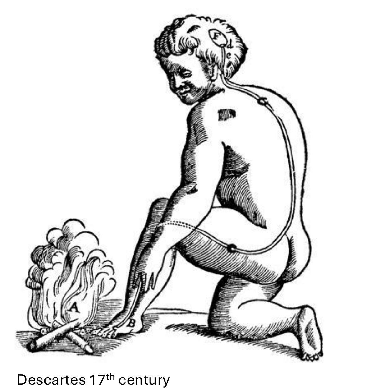
In 1664 René Descartes drew a man kneeling by a fire, a thread running from his foot to his brain. Wrong in almost every detail — right about the one thing that matters: pain is a signal that gets carried somewhere.
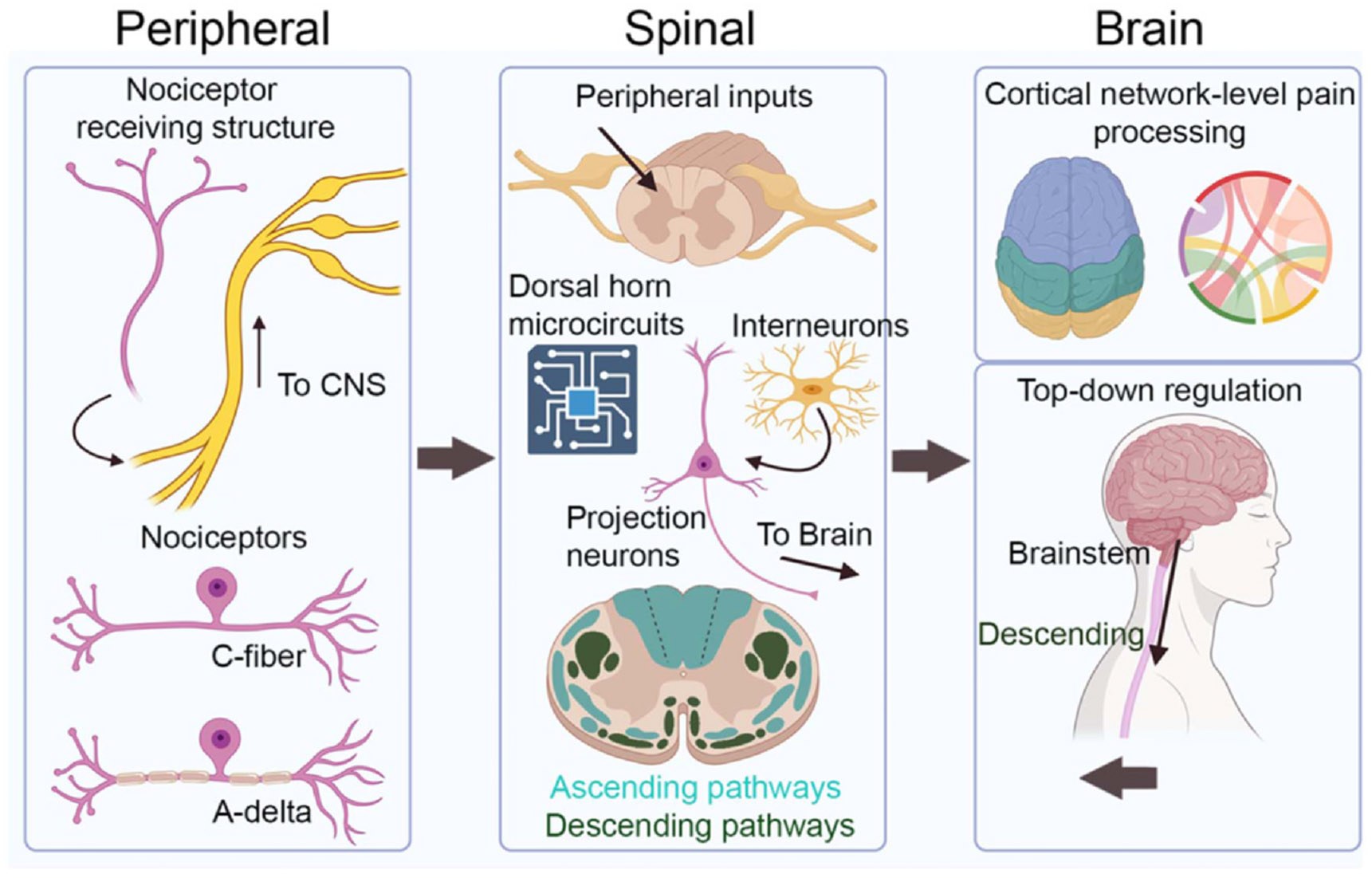
The relay. Nociceptors in the skin detect damaging stimuli and synapse in the dorsal horn. A small population of spinal projection neurons carries that signal to the brainstem and thalamus. Ultimately cortex, where it becomes the thing we call pain.
Why it matters. Changing primary sensory areas changes how organisms behave — and what the animal, or the person, feels.
Pain is not a signal the brain receives. It is a conclusion the brain reaches — and the spinal cord decides what to go on.
The premise of the labStep one: find the neurons
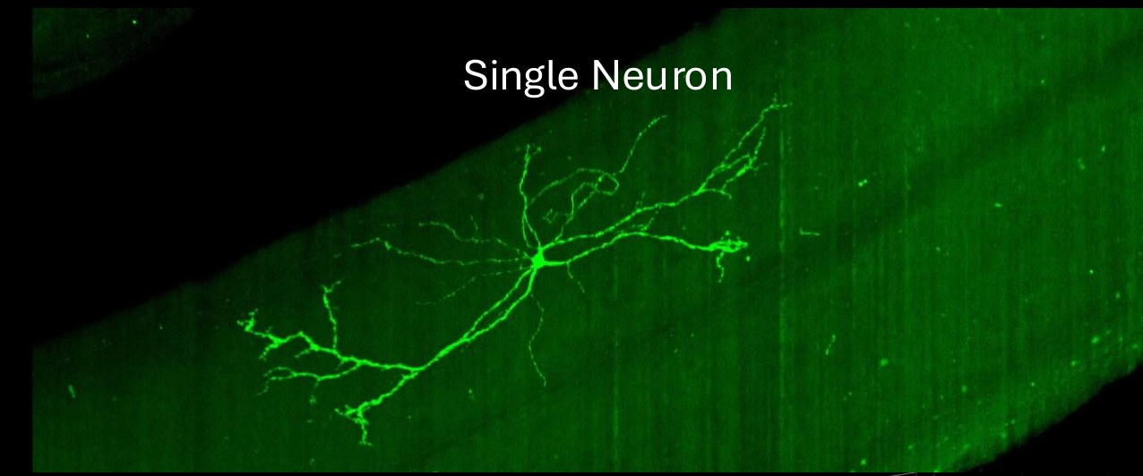
Rare cells. Spinal projection neurons number a few thousand across the whole cord, and until recently there was no clean way to label them as a class. The Phox2a-Cre line changed that (Roome et al.).
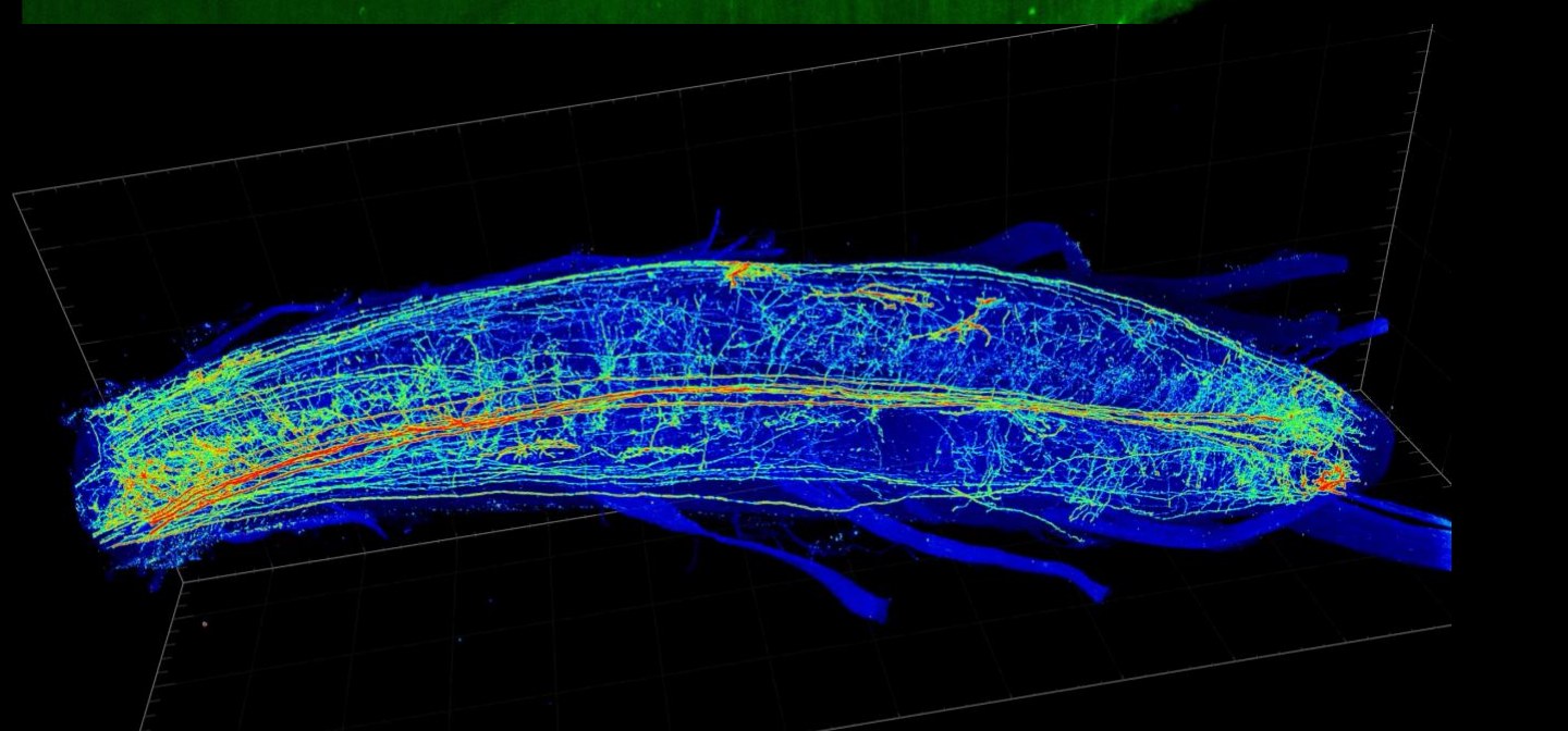
Sparse on purpose. Crossing to a sparse reporter — MORF-GFP, from the Yang lab at UCSD — labels a random handful of cells at a time. With a dense label every arbor overlaps every other; with a sparse one, a single neuron can be reconstructed whole.
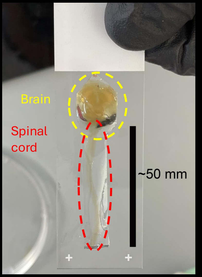
Whole tissue. We clear the entire brain and cord — roughly 50 mm of tissue made transparent — and image it on a light-sheet microscope. The result is an intact three-dimensional map, not a stack of sections.
Step two: record their activity
The hard part. Anatomy tells you what connects to what, not what happens when you pinch a paw. The cord moves with every breath and heartbeat, sits beneath bone and dura, and the surgical window scars over within days.
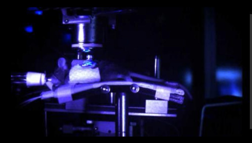
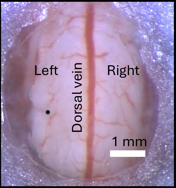
The method. Work we developed at UCSF and published in Nature Methods made long-term optical imaging of the spinal cord in awake, behaving mice possible for the first time: a calcium indicator in projection neurons, a chronic window over the dorsal cord, and an epifluorescence microscope above it.
What we look for. A change in selectivity. In an uninjured animal, populations responding to innocuous touch, noxious mechanical and noxious thermal stimuli overlap only modestly. After nerve injury they converge — cells that should respond only to damage begin responding to a light brush.
Why that matters. Convergence is a plausible circuit-level substrate for allodynia: pain from a stimulus that should not hurt.
Step three: measure the behavior
Beyond thresholds. Classical pain assays measure withdrawal thresholds and latency to lift a paw.
What we do instead. Markerless pose estimation, trained on a few hundred hand-annotated frames and then applied automatically to hours of unlabeled video.
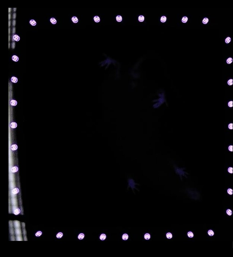
The payoff. A sensitized animal does not simply flinch more. Its whole behavioral repertoire reorganizes — flinching and licking appear where they were nearly absent, grooming rises, exploratory rearing falls away. A single-number threshold assay captures almost none of that.
We want to be able to say: this neuron changed, on this day, and here is what the animal started doing differently.
Why all three levels, in the same animalsWhat we are working on now
The brake. Descending noradrenergic projections from the locus coeruleus actively suppress nociceptive transmission. Duloxetine and related drugs work, when they work, largely by leaning on that system. After nerve injury the brake weakens.
When does it fail? We chart the time course of LC–spinal α2-adrenergic dysfunction rather than sampling a single endpoint, because the therapeutic question is a question about timing.
Does the cause matter? We compare oxaliplatin-induced peripheral neuropathy — bilateral and distal-predominant — against traumatic nerve injury.
Is there a window? If the brake degrades progressively rather than failing at once, there should be a period during which it stays rescuable. Finding that window, and the mechanism that closes it, is the goal.